PEAX Equipment

Is there a (deer) doctor in the house?

Wild Bill

Well-known member
Joined
Dec 9, 2015
Messages
1,504
Location
SD
I shot this buck in North Central SD a couple weeks ago, and while skinning the skull to euro it I discovered an awful stench in the jaw area that was associated with a large infected pus pocket on one side and a smaller on the other, but not as bad.
I figured it was due to fighting and didn’t think much other than it was gross and sucked to take care of.
After an initial few hours in a simmer, I pulled it out to do some fleshing and teeth started falling into the water. Turns out the skull where the pus pockets were is pretty deteriorated and honeycombed, almost.
Just wondering if anyone has any idea what this might be from? It was very localized to the jaw area and the rest of the meat and carcass was perfect, besides a fracture in his maxilla near his nasal cavity, which I’m almost positive would be from fighting.
He was a 4x4 whitetail, probably 3-1/2 years old based on rack and body size.
 

Attachments

  • 03B136C0-C444-477B-8132-F02D8EF6319C.jpeg
    03B136C0-C444-477B-8132-F02D8EF6319C.jpeg
    2.2 MB · Views: 74
  • EAD62733-3166-4A32-8EDD-7B19B7E19756.jpeg
    EAD62733-3166-4A32-8EDD-7B19B7E19756.jpeg
    1.3 MB · Views: 71
  • 2C9BA52E-9758-48F3-8513-68E426F355B6.jpeg
    2C9BA52E-9758-48F3-8513-68E426F355B6.jpeg
    1.4 MB · Views: 60
  • 716B5C3E-1148-4D19-83D9-34EA98E16901.jpeg
    716B5C3E-1148-4D19-83D9-34EA98E16901.jpeg
    1.3 MB · Views: 61
  • 58D0DB52-3444-4F79-9E4E-DEA7ED06F17B.jpeg
    58D0DB52-3444-4F79-9E4E-DEA7ED06F17B.jpeg
    1.6 MB · Views: 61
  • 91C7A6EA-BF62-4273-9584-A3BBC5EE5F31.jpeg
    91C7A6EA-BF62-4273-9584-A3BBC5EE5F31.jpeg
    1.3 MB · Views: 53
  • image.jpg
    image.jpg
    1.6 MB · Views: 70
Last edited:
That’s pretty interesting. Did you notice any swelling, or did he look pretty normal outwardly?

Oral infections aren’t exactly uncommon in cervids. Abrasions from rough forage, or other oral or tooth injuries can get infected. They can get what’s called lumpy jaw, which is an oral infection that often causes swelling that looks like chipmunk cheeks, which is why I ask. Your deer looks like it’s had a more chronic infection to cause that much bone damage, but it’s hard to say exactly what caused it. I’ve never seen one that extensive.

I killed a whitetail once that had a small abscess that had eaten away a lot of the bone and exposed the root of a premolar on one side. He was missing the same tooth on the other side and had bone damage that was much older and healed over. The only one like that I’ve ever shot, that I noticed anyway.
 
Gnarly. I've seen that in people with severe periodontal disease. In people the maxilla is more spongy and porous when compared to the mandible. There is usually a corticated "plate" of bone on the outside of the maxilla, what we refer to as the "buccal plate". In more severe periodontal disease this plate and the bone around the roots can be compromised from bacteria/inflammation and this results in severe bone loss, abscesses, bad odor, and loss of attachment apparatus around the teeth.

You can see where there appears to be a plate of bone covering the roots of the teeth towards the front, but it appears broken off where the molars fell out.

No idea if that's what is occurring here or not, just throwing out an observation which parallels what I see in 2 legged animals.
 
That’s pretty interesting. Did you notice any swelling, or did he look pretty normal outwardly?

Oral infections aren’t exactly uncommon in cervids. Abrasions from rough forage, or other oral or tooth injuries can get infected. They can get what’s called lumpy jaw, which is an oral infection that often causes swelling that looks like chipmunk cheeks, which is why I ask. Your deer looks like it’s had a more chronic infection to cause that much bone damage, but it’s hard to say exactly what caused it. I’ve never seen one that extensive.

I killed a whitetail once that had a small abscess that had eaten away a lot of the bone and exposed the root of a premolar on one side. He was missing the same tooth on the other side and had bone damage that was much older and healed over. The only one like that I’ve ever shot, that I noticed anyway.
Yeah I looked into it prior to the post and saw lumpy jaw but he had no signs of that. He acted and looked as normal as any deer I’d ever shot.
Here’s a pic for reference.
 

Attachments

  • B2D67451-785F-4AB4-896B-8DF235ADFBE9.jpeg
    B2D67451-785F-4AB4-896B-8DF235ADFBE9.jpeg
    6 MB · Views: 54
Yeah I looked into it prior to the post and saw lumpy jaw but he had no signs of that. He acted and looked as normal as any deer I’d ever shot.
Here’s a pic for reference.
I figured. The swelling is more due to soft tissue involvement, usually shorter-term. That kind of infection in the bone must have been there for quite a while so makes sense he wouldn’t have too much outward sign.

Pretty buck though!
 
Your images have the classic hard tissue appearance of periodontal (gum) disease. In humans, this is the biggest reason for tooth loss after the age of 20. Most of that disease process will be painless, right up until it’s too late to save the tooth. Common signs of gum disease include easily bleeding gums, bad breath, loosening teeth, shifting teeth, and oftentimes one can see the buildup of tartar at the gumline.

Bacteria secrete toxins which the host’s immune system will recognize and respond to, migrating to the hard/soft tissue interface, attempting to control these bacterial “invaders”. The problem with this is that the bacteria aren’t actually “in” our body, but instead are living on the tooth, below the gumline in the sulcus (like a moat around a castle) where our immune cells can’t really do a great job.

Your deer‘s molars took the biggest hit. Like humans, those teeth are multi rooted, which provides ample hidey-holes for the bacteria to hide. Some of your photos show significant layers of calculus (“tartar”) which provide the gum disease inducing bacteria another spot to settle and do their dirty work.

Very few deer live longer than 7 years. Most humans don’t show bone loss until they are well into their twenties. With the life expectancy for humans lengthening in the last 100+ years, there has been a significant increase in humans who suffer tooth loss from gum diseases. In my grandparents day, it wasn’t uncommon to remove a diseased tooth instead of trying to rehab it with fillings, root canals, crowns and gum disease treatments.

Vets have discovered a revenue stream and service for their small animal practices. General anesthesia and hygiene care for Fido or Fluffy costs more at the many vet’s office than at my office for similar care (root planing).
 
I’ve seen things similar quite a few times over the year in the taxidermy shop doing skull mounts. Most of what I see are cranial abscess disease and lumpy jaw/tooth abscess on the lower jaw. I’ve seen that kind of upper molar decay but I don’t remember anything as severe as what you’ve got there. Definitely a conversation piece!
 
Yeah I looked into it prior to the post and saw lumpy jaw but he had no signs of that. He acted and looked as normal as any deer I’d ever shot.
Here’s a pic for reference.
C’mon, I think this whole thread was just an excuse for this #humblebrag
Kidding—great buck!
 
Your images have the classic hard tissue appearance of periodontal (gum) disease. In humans, this is the biggest reason for tooth loss after the age of 20. Most of that disease process will be painless, right up until it’s too late to save the tooth. Common signs of gum disease include easily bleeding gums, bad breath, loosening teeth, shifting teeth, and oftentimes one can see the buildup of tartar at the gumline.

Bacteria secrete toxins which the host’s immune system will recognize and respond to, migrating to the hard/soft tissue interface, attempting to control these bacterial “invaders”. The problem with this is that the bacteria aren’t actually “in” our body, but instead are living on the tooth, below the gumline in the sulcus (like a moat around a castle) where our immune cells can’t really do a great job.

Your deer‘s molars took the biggest hit. Like humans, those teeth are multi rooted, which provides ample hidey-holes for the bacteria to hide. Some of your photos show significant layers of calculus (“tartar”) which provide the gum disease inducing bacteria another spot to settle and do their dirty work.

Very few deer live longer than 7 years. Most humans don’t show bone loss until they are well into their twenties. With the life expectancy for humans lengthening in the last 100+ years, there has been a significant increase in humans who suffer tooth loss from gum diseases. In my grandparents day, it wasn’t uncommon to remove a diseased tooth instead of trying to rehab it with fillings, root canals, crowns and gum disease treatments.

Vets have discovered a revenue stream and service for their small animal practices. General anesthesia and hygiene care for Fido or Fluffy costs more at the many vet’s office than at my office for similar care (root planing).
Thanks for the insight! And yes there was significant tartar on the molars when they fell out. some of it cleaned up and fell off while I was working the skull, but there's still some remaining.
 
Your images have the classic hard tissue appearance of periodontal (gum) disease. In humans, this is the biggest reason for tooth loss after the age of 20. Most of that disease process will be painless, right up until it’s too late to save the tooth. Common signs of gum disease include easily bleeding gums, bad breath, loosening teeth, shifting teeth, and oftentimes one can see the buildup of tartar at the gumline.

Bacteria secrete toxins which the host’s immune system will recognize and respond to, migrating to the hard/soft tissue interface, attempting to control these bacterial “invaders”. The problem with this is that the bacteria aren’t actually “in” our body, but instead are living on the tooth, below the gumline in the sulcus (like a moat around a castle) where our immune cells can’t really do a great job.

Your deer‘s molars took the biggest hit. Like humans, those teeth are multi rooted, which provides ample hidey-holes for the bacteria to hide. Some of your photos show significant layers of calculus (“tartar”) which provide the gum disease inducing bacteria another spot to settle and do their dirty work.

Very few deer live longer than 7 years. Most humans don’t show bone loss until they are well into their twenties. With the life expectancy for humans lengthening in the last 100+ years, there has been a significant increase in humans who suffer tooth loss from gum diseases. In my grandparents day, it wasn’t uncommon to remove a diseased tooth instead of trying to rehab it with fillings, root canals, crowns and gum disease treatments.

Vets have discovered a revenue stream and service for their small animal practices. General anesthesia and hygiene care for Fido or Fluffy costs more at the many vet’s office than at my office for similar care (root planing).
I had not really thought about the age issue, but I did initially think of periodontal disease. Could be the aggressive form rather than Chronic but even at 7 years old that would be very extreme for an adult who has had their permanent teeth for a similar amount of time. As I look closer at the bone it looks to be "mottled" or something. It would be interested to see a biopsy for sure. I wonder if deer can get Squamous Cell Carcinoma or other pathologies that Oral and Maxillofacial Surgeons would be familiar with
 
Super interesting. I will defer to people who know more than me on your buck specifically, but I will add for the audience that it's *wild* how many deer run around with abscesses in their foreheads, face, neck etc. without anyone being able to see them from the outside.

Skinning out a couple hundred deer heads a year, I bet 5-10 percent of bucks have green snotty abscesses somewhere under their head/face skin, and 1-2 percent carry broken antler tips under their skin. Talk about an absess.
 
I shot this buck in North Central SD a couple weeks ago, and while skinning the skull to euro it I discovered an awful stench in the jaw area that was associated with a large infected pus pocket on one side and a smaller on the other, but not as bad.
I figured it was due to fighting and didn’t think much other than it was gross and sucked to take care of.
After an initial few hours in a simmer, I pulled it out to do some fleshing and teeth started falling into the water. Turns out the skull where the pus pockets were is pretty deteriorated and honeycombed, almost.
Just wondering if anyone has any idea what this might be from? It was very localized to the jaw area and the rest of the meat and carcass was perfect, besides a fracture in his maxilla near his nasal cavity, which I’m almost positive would be from fighting.
He was a 4x4 whitetail, probably 3-1/2 years old based on rack and body size.
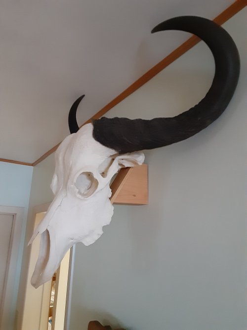
Obviously gum disease. Where did you shoot this buck? Looks like he's been on a diet that was rich in sweets. Someone in the neighborhood putting rich crap in a feeder? Apples? Here's the first buffalo I shot in Africa. The taxidermist there cooked all the teeth out. Not uncommon but they should have replaced them. Note that this buff had been missing a molar for some time as the gum is recessed. At twenty-five years old I'm surprised it had any teeth left.20201008_114835.jpg
 
Last edited:
I have seen puss pockets in the joints of the front and rear quarters. The deer had a smell when I walked up to them. It was associated with being hit by a car and managing to live, that being said the infection would have likely killed them over time or left them crippled enough for coyotes to get a dinner.

Looking at the pictures closer I would say impact from hitting something or getting hit by something and resulting in a fractured jaw.
 
Advertisement

Latest posts

Forum statistics

Threads
114,369
Messages
2,054,463
Members
36,570
Latest member
ihgodfather
Back
Top